
What is it?
There are two distinct conditions referred to as Köhler’s disease, both affecting the bones of the foot. They are known as Köhler I and Köhler II (also called Freiberg’s disease), and they belong to the group of osteochondroses—disorders that affect growing bones in children and adolescents.
- Köhler I is an osteochondrosis of the navicular bone in the midfoot. It is relatively rare, usually unilateral, and most often diagnosed in boys between the ages of 3 and 8.
- Köhler II (Freiberg’s disease) is an osteochondrosis of the proximal epiphysis of the second metatarsal. It primarily affects females between the ages of 18 and 30, though it can also appear during adolescence.
Causes of Köhler’s Disease
The exact cause of Köhler’s disease is still uncertain. A widely accepted theory suggests that mechanical stress during weight-bearing activities (such as running and jumping) leads to a vascular deficit, reducing blood supply to the bone. This ischemia may result in bone tissue necrosis and collapse of the navicular bone, which appears flattened on imaging.
Blood flow to the bone is strongly influenced by repetitive loading, impact, and rotational stresses. These factors may delay normal ossification and interfere with healthy bone development.
Diagnosis
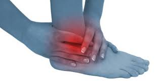
Köhler’s disease can sometimes be asymptomatic. When symptoms occur, they typically include:
- Swelling (edema) on the inner dorsal side of the foot
- Pain in the plantar region, which worsens after prolonged standing or when wearing high-heeled shoes
- Functional limitations, including difficulty walking
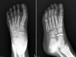
Diagnosis is based on clinical evaluation by a specialist, medical history, and X-rays. Radiographs usually show a flattened navicular bone with areas of sclerosis and fragmentation. For thorough assessment, imaging of both feet is recommended.
Treatment
The disease course is typically benign, with complete recovery expected within 18 to 24 months.
During the 2–3 months when pain is most acute, limiting weight-bearing activities is advised. After the acute phase, the use of orthotic insoles may be recommended. By supporting the longitudinal arch of the foot, orthotics help reduce stress on the navicular bone and allow a gradual return to physical activity.
The Riva Method and Köhler’s Disease
In children over the age of six, the Riva Method can be particularly beneficial. It provides specific categories of medium-to-low intensity mechanical stimulation, which are essential for promoting structural remodeling of the foot’s musculoskeletal system.
Through proprioceptive training and controlled load distribution, the Riva Method supports natural bone healing, enhances functional stability, and contributes to a safer, more effective recovery.